Mon–Sat: 9:30 AM – 7:30 PM  ·  Sun: By Appointment Only
Seek care promptly

Wisdom Tooth Pain & Impaction

Pain or swelling behind the last molar deserves a proper examination. We identify the cause, explain whether monitoring or treatment is appropriate, and plan the next step with you.
Urgent: Pain with swelling or fever needs prompt examination. Difficulty breathing, speaking or swallowing, extensive mouth swelling, or swelling around the eye requires immediate emergency medical care.
Signs & symptoms

What You May Notice

These symptoms can have several causes. An examination helps establish whether a wisdom tooth is responsible and whether treatment is needed.

Find what feels familiar.
Tap a symptom to read more.

Discomfort behind the last visible molar, which may be intermittent or persistent.

Redness or soreness around a partly erupted tooth can indicate local inflammation.

A partly erupted or angled tooth may create an area that is difficult to clean.

Food and plaque beneath a gum flap may contribute to a bad taste or odour.

Jaw stiffness or pain on opening needs examination, especially when it is increasing.

Wisdom-tooth pain can be felt elsewhere, but jaw-joint, muscle and ear causes must also be considered.

Some signs need prompt attention

Cheek or jaw swelling: Swelling with pain, fever or increasing limitation requires prompt attention.

Fever or swallowing pain: These can indicate spreading infection and should not be ignored.

Immediate emergency medical care: Difficulty breathing, speaking or swallowing, extensive mouth swelling, or swelling around the eye. Do not wait for a clinic appointment.

Interactive wisdom tooth guide

Understand Your Wisdom Tooth and the Next Step

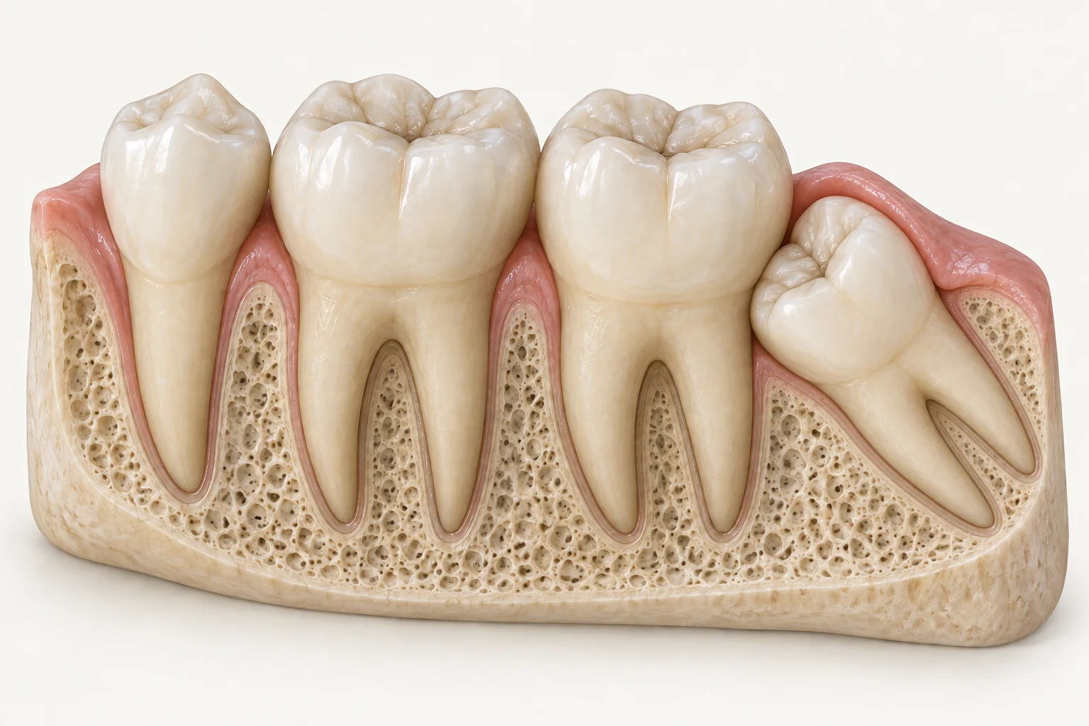
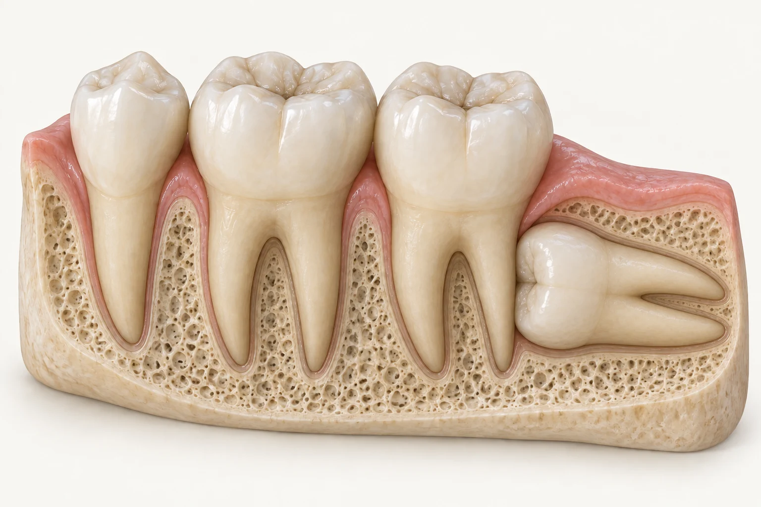
Explore four example positions using the same neighbouring teeth for context. The illustrations explain anatomy; your dentist uses examination and appropriate imaging to advise on your tooth.

One view. A clearer explanation.
Choose a position to explore the tooth, what we check and the possible next steps.
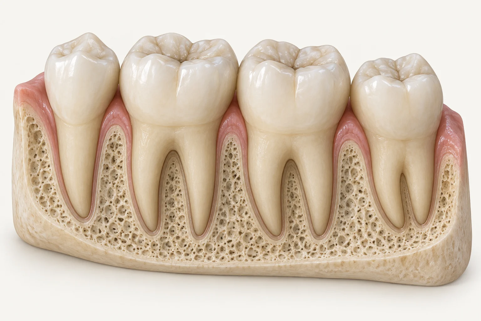
4 teeth · lower-jaw view
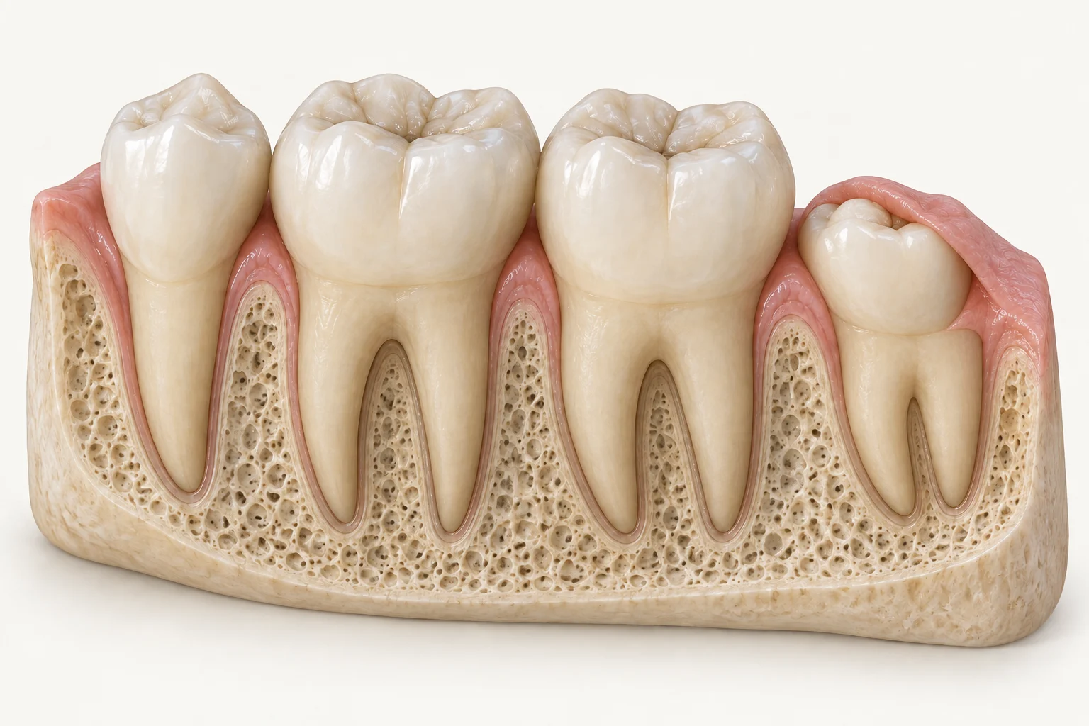
Illustrative lower-jaw cutaway with a premolar, two neighbouring molars and a fully erupted wisdom tooth.
Second
premolar
First
molar
Second
molar
Wisdom tooth
Third molar
Explore possible problems

Choose an area to see why it may need checking.

The image shows an example position, not a diagnosis.

EXAMPLE 01

Fully erupted

What you are seeing

The last tooth is upright, with its crown above the gum. The three teeth beside it provide context.

What the dentist checks

Cleaning access, gum health, decay and the way the teeth meet.

What may happen next

If it is healthy and can be maintained, review may be appropriate. Any disease needs to be investigated; eruption alone does not decide treatment.
Position alone does not decide removal. Examination, imaging when indicated, your health and a discussion of benefits and risks guide the plan.

Could the pain come from somewhere else?

Yes. A neighbouring tooth, jaw muscles, the jaw joint or an ear-related problem may need examination. Finding a wisdom tooth on an X-ray does not by itself establish the cause of pain.

Explore orofacial pain consultation →
What Happens at the Visit?
  1. Listen and examine. We ask about your symptoms and health, then examine the tooth, gum, adjacent molar and jaw.
  2. Image when needed. The choice of imaging depends on what needs clarification; not everyone needs the same scan.
  3. Discuss the options. These may include local care, review, removal or specialist consultation. They are possible routes, not steps everyone must follow.

Antibiotics are used only when clinically indicated. If removal is recommended, we explain anaesthesia, relevant risks, alternatives and recovery before you decide.
Four-tooth view
Second
premolar
First
molar
Second
molar
Wisdom tooth
Third molar

Illustrative cutaway. The outer bone is opened to reveal the tooth roots; this is not a patient photograph or scan.

After removal

Recovery: What to Expect

Recovery differs with the tooth, the procedure, your health and the anaesthesia used. Follow the instructions provided for your specific treatment.

Protect the healing area

Avoid disturbing the socket and follow the cleaning instructions given by your clinician.

Choose comfortable foods

Soft, cool or lukewarm foods are commonly easier initially. Individual advice may differ.

Use medicines as directed

Take prescribed medicines exactly as advised. Do not start antibiotics independently.

Know when to call

Seek advice for persistent bleeding, rapidly increasing swelling or pain that becomes severe or worsens.
Care and risk reduction

Keeping the Back of the Mouth Clean

Indian woman holding a toothpaste-loaded toothbrush just before brushing in a bathroom

Clean gently and thoroughly

Use a soft toothbrush and the cleaning method recommended for the partly erupted area.

Do not ignore recurrence

Repeated pain, swelling or a bad taste deserves reassessment even if it settles temporarily.

Attend recommended reviews

Review intervals and imaging should be based on the individual tooth and its risks.

Avoid self-medicating

Painkillers and antibiotics can mask symptoms without correcting the underlying problem.

When to Seek Care

Arrange an examination for recurrent pain, tender gum, bad taste, swelling, fever or increasing difficulty opening the mouth.

Prompt dental consultation

Pain with swelling or fever, recurrent inflammation, or increasing limitation in opening the mouth.

Immediate emergency medical care

Difficulty breathing, speaking or swallowing, extensive mouth swelling, or swelling around the eye. Do not wait for a clinic appointment.

Frequently Asked Questions

General information to support your discussion with the treating clinician.
Pain from a wisdom tooth is often felt behind the last visible molar and may occur with tender gum, a bad taste, swelling or difficulty opening the mouth. Similar symptoms can have other causes, so an examination is needed.
Not everyone needs the same imaging. A panoramic X-ray may be recommended when the position of the tooth, nearby structures or disease cannot be assessed fully by examination alone. CBCT is reserved for selected cases.
No. A tooth without symptoms, disease or significant risk may be monitored. The decision depends on the examination, imaging when indicated, your health and the balance of benefits and risks.
Local anaesthesia is used to control pain during treatment. Pressure or movement may still be felt. Your clinician will explain the anaesthesia plan and what to expect for your particular tooth.
Sometimes, but not automatically. The number treated at one time depends on their position, symptoms, complexity, medical factors and the planned anaesthesia.
Your clinician will give procedure-specific advice. Soft, cool or lukewarm foods are commonly easier initially. Avoid disturbing the healing area and follow the instructions provided to you.
Dry socket is a painful healing complication that can occur after extraction when the protective clot is lost or breaks down. Pain that becomes severe or worsens after initially improving needs prompt dental advice.
Some swelling can occur after removal. Seek advice for swelling that is rapidly increasing, accompanied by fever, severe worsening pain or persistent bleeding. Difficulty breathing, speaking or swallowing requires immediate emergency medical care.
Certain positions can increase the risk of decay, gum problems or damage affecting the adjacent second molar. Examination and appropriate imaging help assess that risk.
Recovery varies with the tooth, procedure, anaesthesia and the type of work you do. Ask your treating clinician for an individual recommendation rather than relying on a fixed timeline.

Understand the Findings Before Deciding

Book a consultation with the GardenCity Smiles™ clinical team in Yelahanka. We will explain the findings, suitable options and the next step clearly.
Scroll to Top